Calcification and mass abnormalities in breast mammogram scans
Download scientific diagram | Calcification and mass abnormalities in breast mammogram scans. The calcification distribution depicts tiny flecks of calcium as small white regions on the left side, while the mass is shown as a smooth, well-defined border on the right side. from publication: Multi-Graph Convolutional Neural Network for Breast Cancer Multi-Task Classification | Mammography is a popular diagnostic imaging procedure for detecting breast cancer at an early stage. Various deep learning (DL) approaches to breast cancer detection incur high costs and are prone to classify incorrectly. Therefore, they are not sufficiently reliable to | Breast Cancer, Convolution and Classification | ResearchGate, the professional network for scientists.
Breast Cancer Ultrasonography: Practice Essentials, Role of Ultrasonography in Screening, Breast Imaging Reporting and Data System

Early detection and classification of abnormality in prior mammograms using image-to-image translation and YOLO techniques - ScienceDirect

Segmenting Abnormalities in Mammograms (Part 1 of 3), by Cleon W

Contrast enhanced mammography: focus on frequently encountered benign and malignant diagnoses, Cancer Imaging
Atlas of breast cancer early detection

a) The cropping breast profile image of mdb111 for left MLO
Algorithms, Free Full-Text
:max_bytes(150000):strip_icc()/why-not-annual-ultrasounds-instead-of-mammograms-430185-v2-dd947cd85bdc40a0ad79d4f4761d61b7.png)
Breast Ultrasound vs. Mammography: Which Is Best?

Brendan JENNINGS, Head of Graduate Studies
Mammography report: The abnormal Mammogram - Moose and Doc
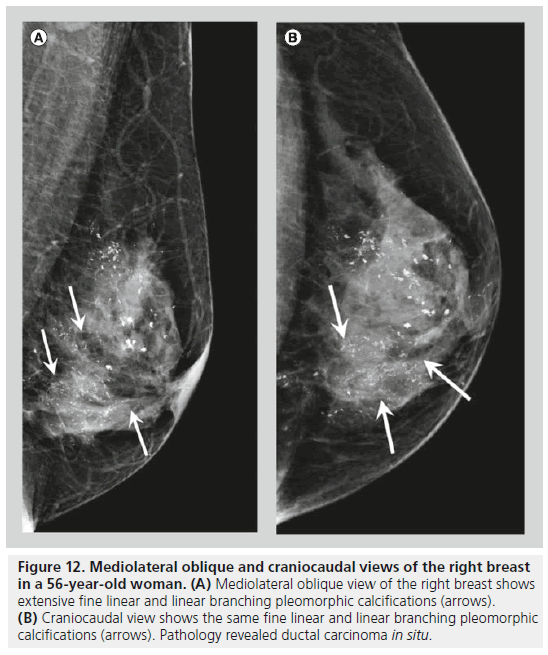
Mammography of breast calcifications
Mimickers of breast malignancy: imaging findings, pathologic concordance and clinical management, Insights into Imaging