Optical microscope images, (a) 0h, (b) 16h, (c) 26h, (d) 38h and (e) 48h

Biological study of skin wound treated with Alginate/Carboxymethyl cellulose/chorion membrane, diopside nanoparticles, and Botox A

Recent advances in optical imaging through deep tissue: imaging probes and techniques, Biomaterials Research
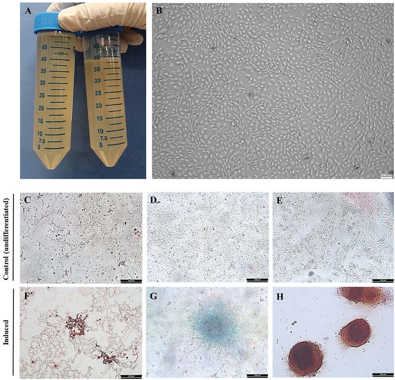
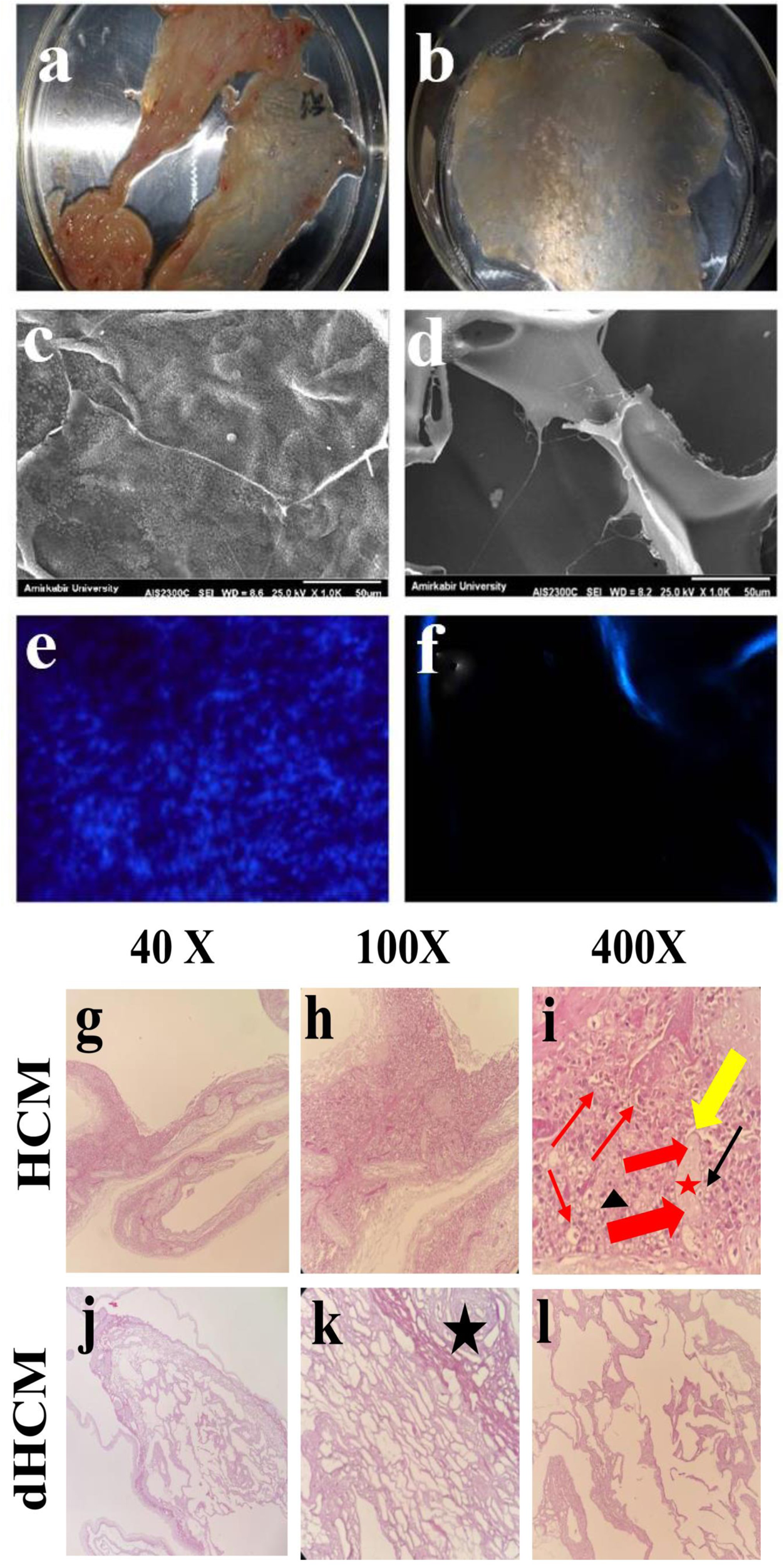
Frontiers Evaluation of stability and safety of equine mesenchymal stem cells derived from amniotic fluid for clinical application
IJMS, Free Full-Text
Recent advances in optical imaging through deep tissue: imaging probes and techniques, Biomaterials Research

Dual mass spectrometry imaging and spatial metabolomics to investigate the metabolism and nephrotoxicity of nitidine chloride - ScienceDirect

Pre-incubation of corneal donor tissue with sCD83 improves graft survival via the induction of alternatively activated macrophages and tolerogenic dendritic cells - American Journal of Transplantation

Optical microscopy (left) and scanning electron microscopy (right)

Optical microscope images, (a) 0h, (b) 16h, (c) 26h, (d) 38h and
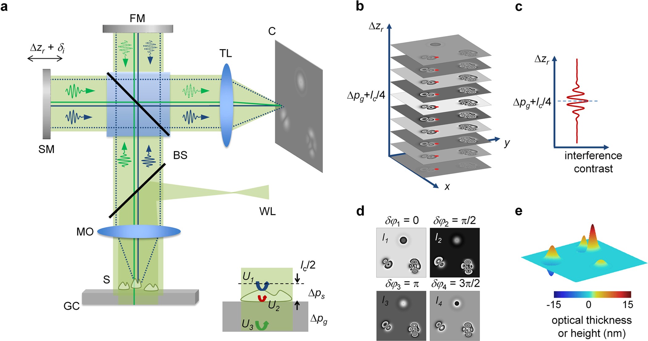
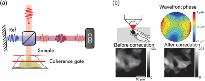
Quantitative reflection phase mesoscopy by remote coherence tuning of phase-shift interference patterns

Motic Panthera C2 Binocular Compound Microscope With Phase Contrast And Darkfield Sliders For Life Science - Microscopy Supplies - Stellar Scientific - Microscopy and Pathology Equipment - Stellar Scientific

PDF) Homogenization heat treatment influence on microstructure

Nanobubble-actuated ultrasound neuromodulation for selectively shaping behavior in mice

Optical microscope and SEM images for determination of fibre

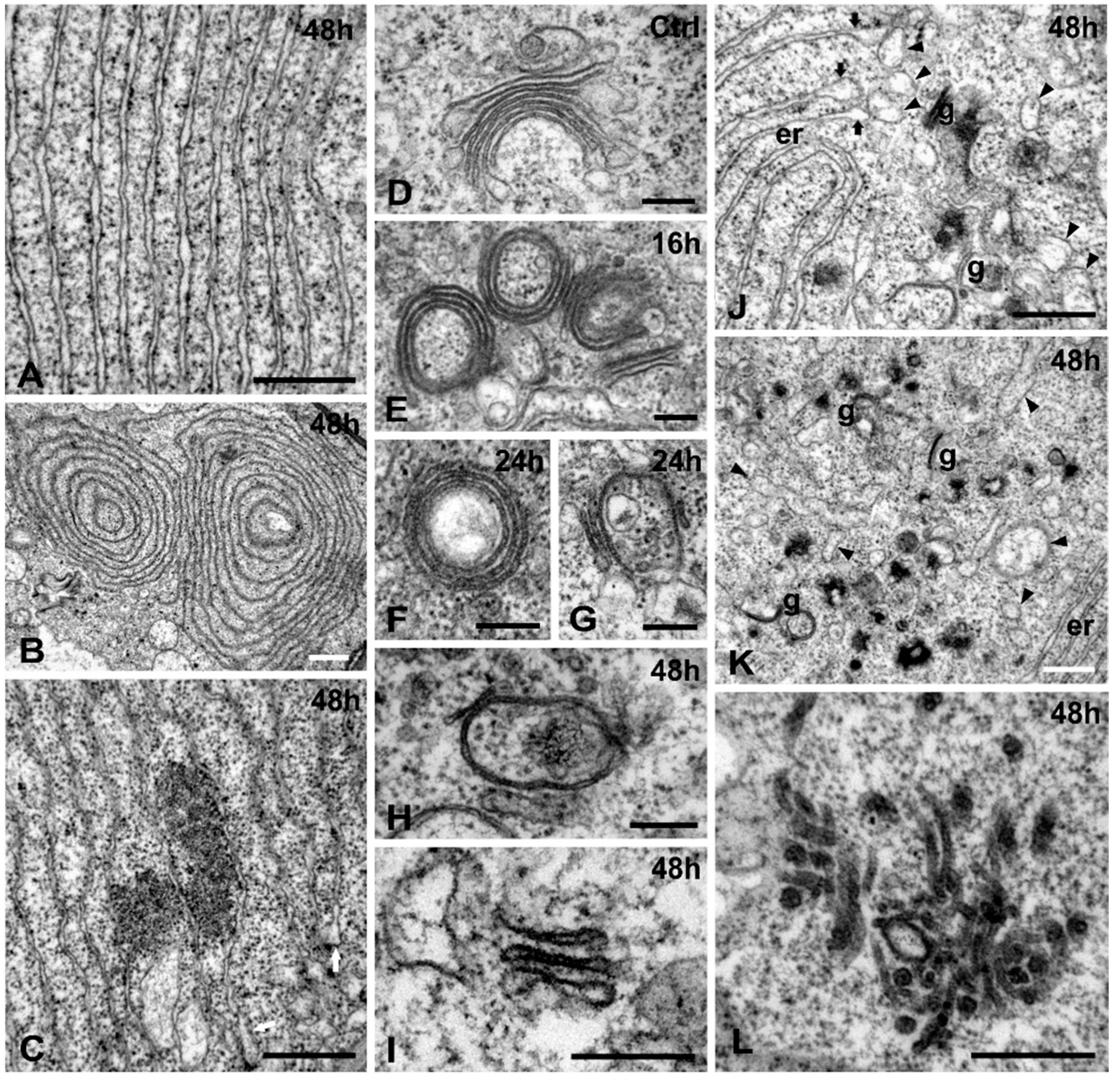
Scanning electron microscope observation of cells incubated for 48