Preoperative tumor size measurement in breast cancer patients: which threshold is appropriate on computer-aided detection for breast MRI?, Cancer Imaging
Background Computer-aided detection (CAD) can detect breast lesions by using an enhancement threshold. Threshold means the percentage of increased signal intensity in post-contrast imaging compared to precontrast imaging. If the pixel value of the enhanced tumor increases above the set threshold, CAD provides the size of the tumor, which is calculated differently depending on the set threshold. Therefore, CAD requires the accurate setting of thresholds. We aimed to compare the diagnostic accuracy of tumor size measurement using MRI and CAD with 3 most commonly used thresholds and to identify which threshold is appropriate on CAD in breast cancer patients. Methods A total of 130 patients with breast cancers (80 invasive cancers and 50 ductal carcinoma in situ [DCIS]) who underwent preoperative MRI with CAD and surgical treatment were included. Tumor size was manually measured on first contrast-enhanced MRI and acquired by CAD using 3 different thresholds (30, 50, and 100%) for each tumor. Tumor size measurements using MRI and CAD were compared with pathological sizes using Spearman correlation analysis. For comparison of size discrepancy between imaging and pathology, concordance was defined as estimation of size by imaging within 5 mm of the pathological size. Concordance rates were compared using Chi-square test. Results For both invasive cancers and DCIS, correlation coefficient rho (r) between tumor size on imaging and pathology was highest at CAD with 30% threshold, followed by MRI, CAD with 50% threshold, and CAD with 100% threshold (all p < 0.05). For invasive cancers, the concordance rate of 72.5% at CAD with 30% threshold showed no difference with that of 62.5% at MRI (p = 0.213). For DCIS, the concordance rate of 30.0% at CAD with 30% threshold showed no difference with that of 36.0% at MRI (p = 0.699). Compared to MRI, higher risk of underestimation was noted when using CAD with 50% or 100% threshold for invasive cancers and when using CAD with 100% threshold for DCIS. Conclusion For CAD analysis, 30% threshold is the most appropriate threshold whose accuracy is comparable to manual measurement on MRI for tumor size measurement. However, clinicians should be aware of the higher risk of underestimation when using CAD with 50% threshold for tumor staging in invasive cancers.

Breast MRI: State of the Art
PDF) Preoperative tumor size measurement in breast cancer patients: which threshold is appropriate on computer-aided detection for breast MRI?
Pancreas: Artificial Intelligence Imaging Pearls - Educational Tools, CT Scanning, CT Imaging
Expert tumor annotations and radiomics for locally advanced breast cancer in DCE-MRI for ACRIN 6657/I-SPY1

PSOWNNs-CNN: A Computational Radiology for Breast Cancer Diagnosis Improvement Based on Image Processing Using Machine Learning Methods

MRI, Clinical Examination, and Mammography for Preoperative Assessment of Residual Disease and Pathologic Complete Response After Neoadjuvant Chemotherapy for Breast Cancer: ACRIN 6657 Trial

Preoperative tumor size measurement in breast cancer patients: which threshold is appropriate on computer-aided detection for breast MRI?, Cancer Imaging
Frontiers The road to breast cancer screening with diffusion MRI

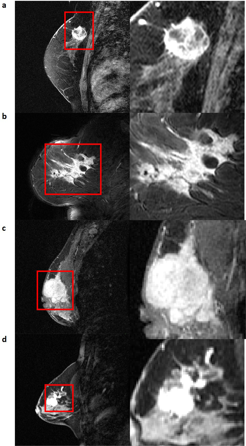
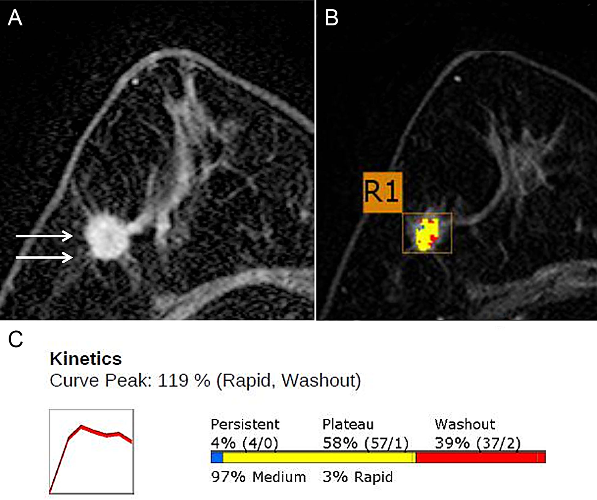
MR images of a 54-year-old-woman with ductal carcinoma in situ. a Axial
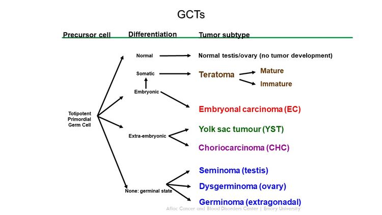
Childhood Extracranial Germ Cell Tumors Treatment (PDQ®) (Health professionals)

Ultrafast dynamic contrast-enhanced breast MRI: association with pathologic complete response in neoadjuvant treatment of breast cancer

Exploration of tumor size measurement methods in preoperative breast cancer assessment using whole-body silicon photomultiplier PET: feasibility and first results

MRI can accurately diagnose breast cancer during lactation

Figure 3 from Can breast MRI computer-aided detection (CAD) improve radiologist accuracy for lesions detected at MRI screening and recommended for biopsy in a high-risk population?