Thoracic spine x-ray
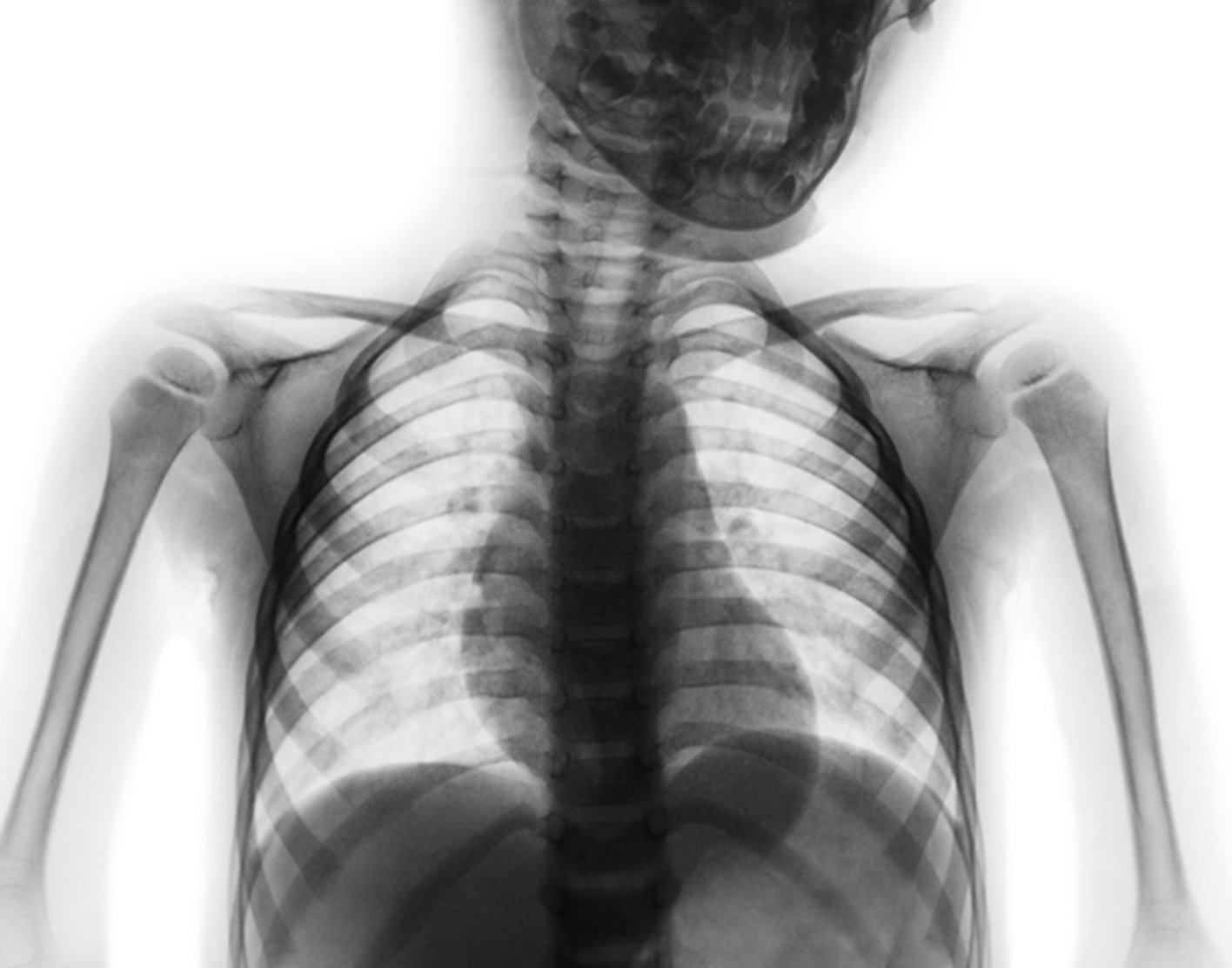
A thoracic spine x-ray is an x-ray of the twelve chest (thoracic) bones (vertebrae). The vertebrae are separated by flat pads of cartilage called disks.
Imaging Anatomy

Thoracic Spine Radiology Tutorial
AP Thoracic Spine X-ray Quiz

Functional Radiographic Analysis of Thoracic Spine Extension Motion in Asymptomatic Men - ScienceDirect

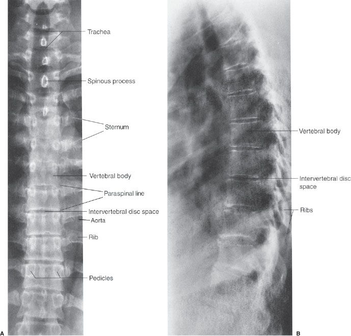
Introduction to Spine Radiographs

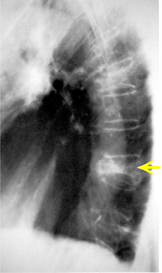
Thoracic Spine Compression Fractures – Vertebra Plana –
9 Spine and Pelvis

Severe thoracic spinal fracture-dislocation without neurological symptoms and costal fractures: a case report and review of the literature, Journal of Medical Case Reports

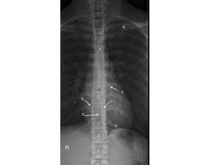
Normal AP thoracic spine radiograph, Radiology Case

Thoracic spine X-rays showing the T7 fracture

Thoracolumbar spine x-rays - Don't Forget the Bubbles

Learning Radiology - Spine Sign

Normal Humans Thoracic-lumbar Spine Stock Image - Image of healthcare, black: 38476127
Plain X-ray Spine