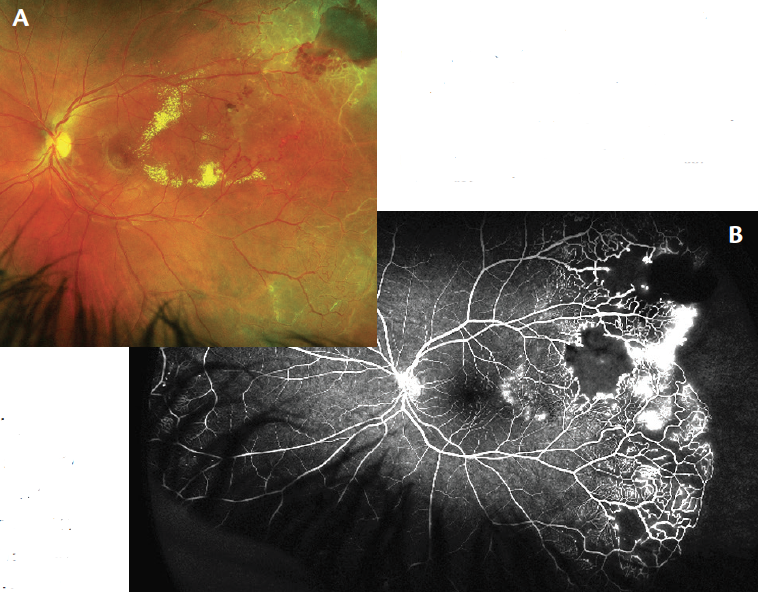
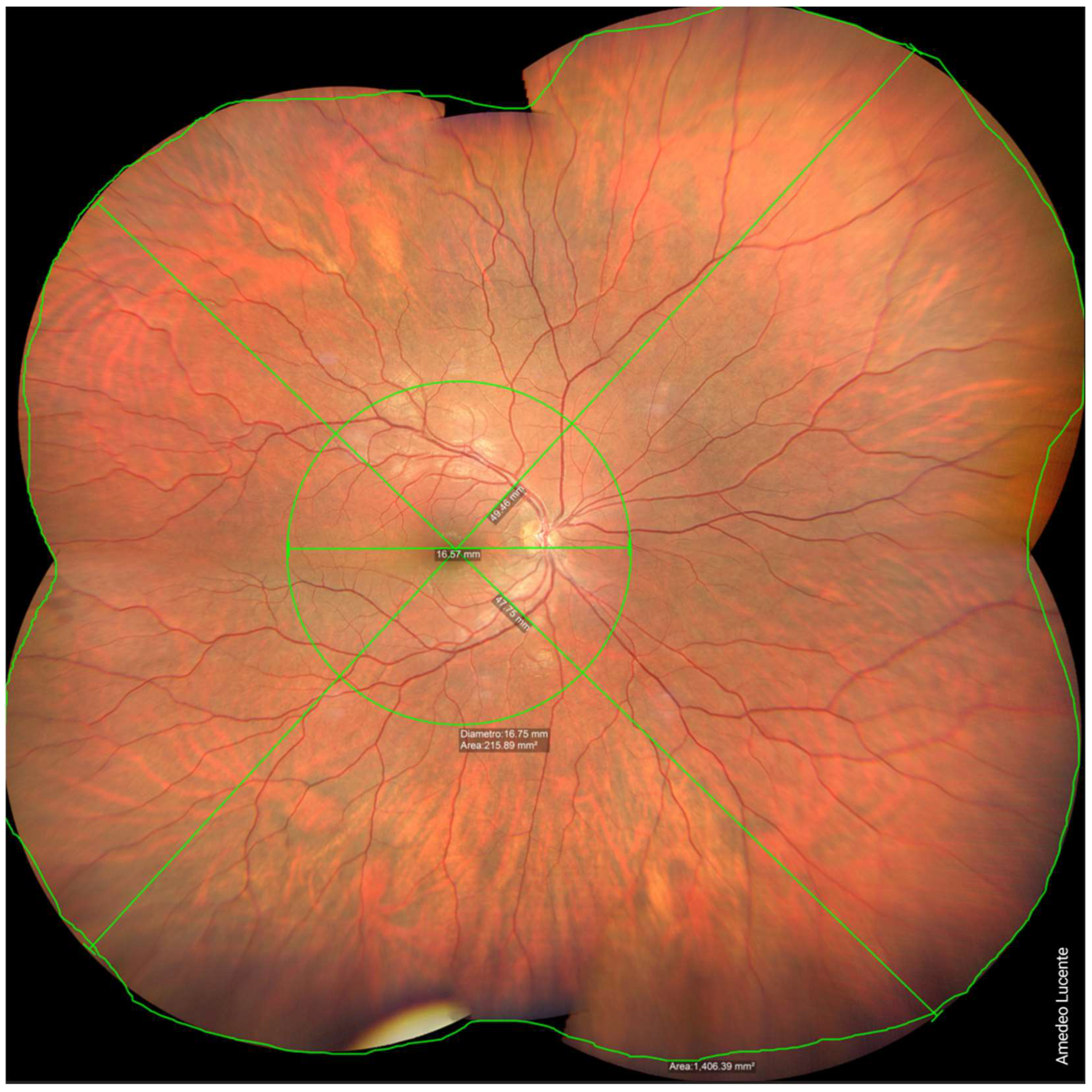
Ultra-wide-field fundus photographs and ultra-wide-field

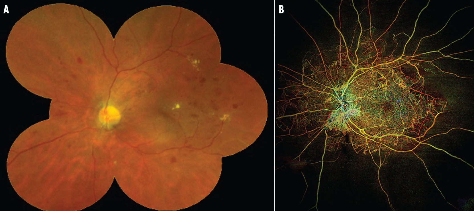
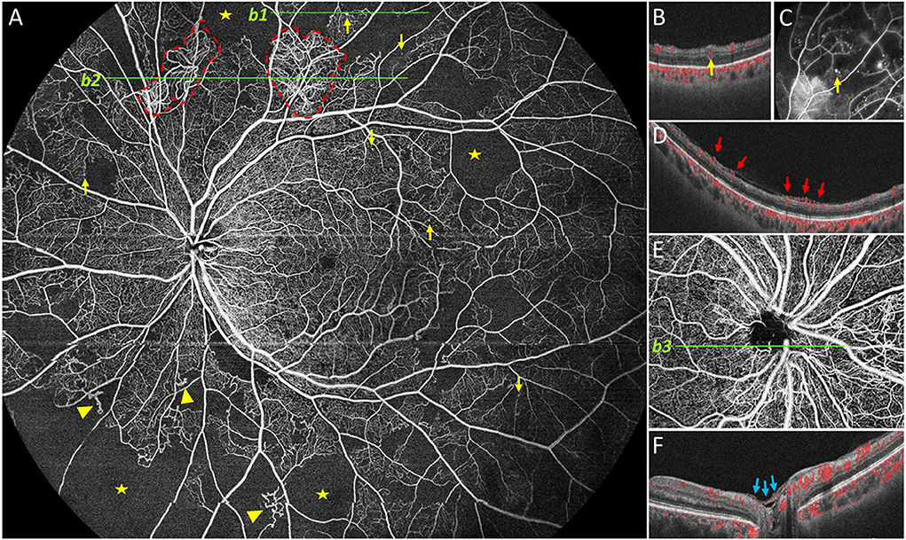
Download scientific diagram | Ultra-wide-field fundus photographs and ultra-wide-field fluorescein angiographic imaging of ocular toxocariasis. (A) A granuloma with mild vitreous opacity. (B) A tractional retinal fold with localized tractional retinal detachment. (C) Diffuse peripheral vascular leakage. (D) A prominent optic disc leakage. from publication: The Clinical Characteristics of Ocular Toxocariasis in Jeju Island Using Ultra-wide-field Fundus Photography | Toxocariasis, Ocular and Photography | ResearchGate, the professional network for scientists.
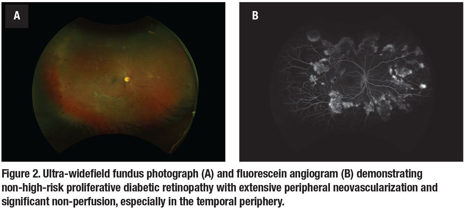
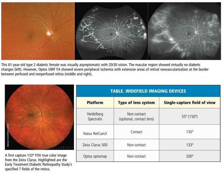
How ultra-widefield imaging is changing our view of DR
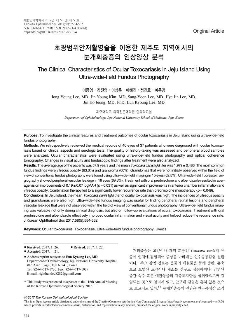
PDF) The Clinical Characteristics of Ocular Toxocariasis in Jeju Island Using Ultra-wide-field Fundus Photography

PDF) The Clinical Characteristics of Ocular Toxocariasis in Jeju Island Using Ultra-wide-field Fundus Photography

Jong Young Lee's research works Jeju National University Hospital, Jeju City and other places
Ultra-Widefield Imaging & Numerous Retinal Pathologies
Ultra-Widefield Imaging Guides Coats Disease Treatment - Retina Today
Life, Free Full-Text
Optomap Ultra Widefield Retinal Imaging

Eun Kyoung Lee's research works Dongguk University, Seoul and other places

PDF) The Clinical Characteristics of Ocular Toxocariasis in Jeju Island Using Ultra-wide-field Fundus Photography

Ultra-Widefield Retinal Imaging, Noosa Optical
Widefield OCTA: A New Way to Stage Diabetic Retinopathy - Retina Today

Jong Young Lee's research works Jeju National University Hospital, Jeju City and other places
Frontiers Ultra-widefield color fundus photography combined with high-speed ultra-widefield swept-source optical coherence tomography angiography for non-invasive detection of lesions in diabetic retinopathy

Sang-Yoon Lee's research works Gachon University, Seongnam-si (kyungwon) and other places