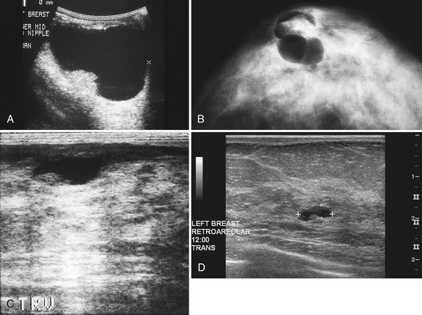
b. Left breast USG showing oval, well-defined, mixed echogenic

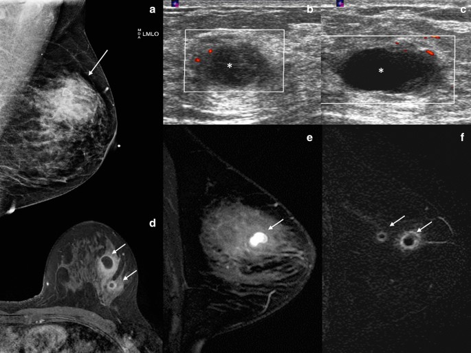
Mimickers of breast malignancy: imaging findings, pathologic

How to Use Breast Ultrasound

Hyperechoic breast images: all that glitters is not gold

c. Left breast USG-irregular, ill-defined, multilobulated

Solid masses: What are the underlying histopathological lesions

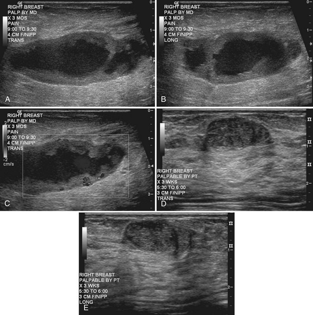
a. Case 1. Right breast USG showing well-defined, hypoechoic mass

PDF) Tumoral pseudoangiomatous stromal hyperplasia: Radiological

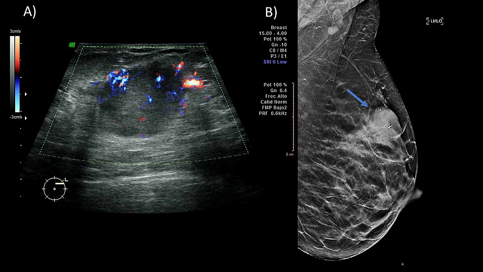
A) Mammography of left breast shows a well-defined, oval shaped

Imaging of fat‐containing lesions of the breast: A pictorial essay

Ultrasonography
Breast Ultrasound
Cureus Pregnancy-Associated Breast Cancer: What Radiologists
Breast Ultrasound