Microcalcification distribution over the smallest breast volume (d =

What does breast DCIS look like?

Serge MULLER, Scientific Advisor, PhD, HDR, Institut de Cancérologie Gustave Roussy, Villejuif, IGR, Department of Medical Imaging

Growth Dynamics of Mammographic Calcifications: Differentiating Ductal Carcinoma in Situ from Benign Breast Disease

Anna MIRA, Research Associate, Doctor of Engineering, King's College London, London, KCL, Division of Imaging Sciences and Biomedical Engineering

Pablo MILIONI DE CARVALHO, Senior Researcher, PhD, General Electric, CA, GE, Division of Healthcare Technology
Electronics, Free Full-Text
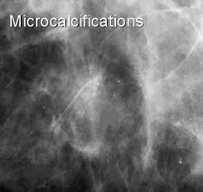
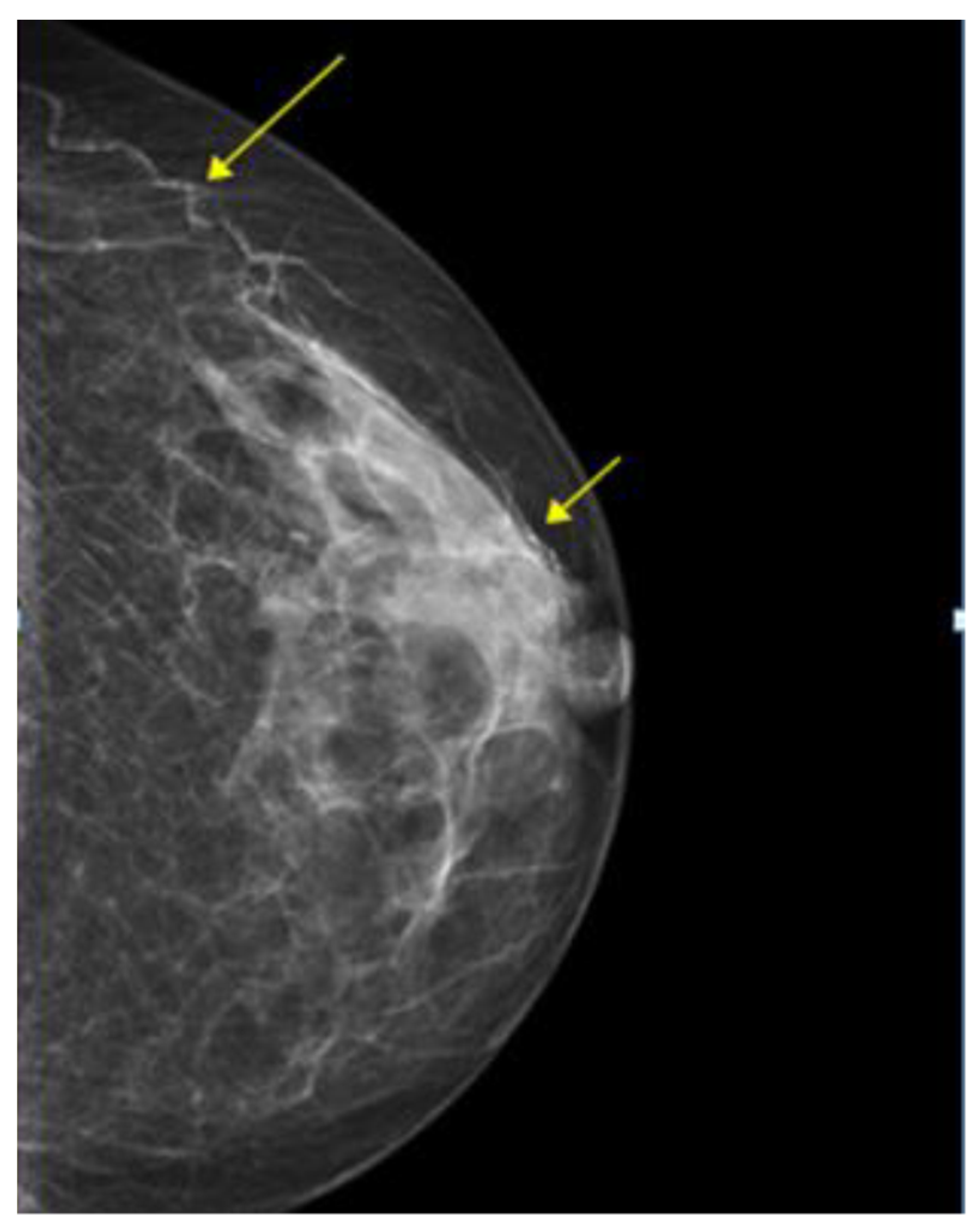
Mammography of breast calcifications

PPT - Breast Calcifications - Differential diagnosis and BIRADS PowerPoint Presentation - ID:3713492

Overview of the processing steps: (1) determination of 3D breast volume

Microcalcifications in Breast Cancer